French national reference center for craniostenoses and craniofacial malformations (CRANIOST)
![]()
![]() Craniofacial surgery unit at Necker-Enfants malades hospital is an internationally renowned reference center. The work of the founders of the center, professors Renier and Marchac, have allowed to build the bases of current clinical and surgical management of these pathologies and to develop the research to understand their genetic causes.
Craniofacial surgery unit at Necker-Enfants malades hospital is an internationally renowned reference center. The work of the founders of the center, professors Renier and Marchac, have allowed to build the bases of current clinical and surgical management of these pathologies and to develop the research to understand their genetic causes.
Since its certification in 2006, the team has paid particular attention to multidisciplinary craniofacial care, associating in particular the craniofacial plastic surgery and pediatric neurosurgery teams.
The craniofacial surgery activity of the Necker-Enfants malades hospital is located within the pediatric neurosurgery department. It is a reconstructive surgery activity for neurosurgical complications.
One of the largest cohorts in the world (4,500 craniostenoses operated on) is followed in the unit, with more than 200 operations per year on an active file of 1,270 patients, with approximately 370 new patients. Approximately 1,200 consultations for these patients are provided each year, with approximately 35% of patients from outside the region and 15% of foreign patients. The number of annual interventions is constantly increasing, due to recruitment and iterative interventions on certain patients.
![]() This reference center is affiliated with the TETECOU rare diseases healthcare network and
This reference center is affiliated with the TETECOU rare diseases healthcare network and ![]() the CRANIO European reference network (ERN) on craniofacial anomalies and ENT disorders.
the CRANIO European reference network (ERN) on craniofacial anomalies and ENT disorders.
Medical
team

Medical leader
Dr Giovanna Paternoster
MD
Medical consultant
Dr Eric Arnaud
MD
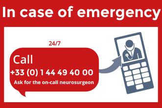
In case of emergency
The missions of the center are:
- to establish a diagnosis with certainty thanks to a specialized team;
- to offer personalized treatment based on multidisciplinary care;
- to train the parents of patients and their families;
- to prevent complications;
- to ensure continuity of care until adulthood;
- to develop clinical and fundamental research programs at national and international level;
- to study the pathophysiological and etiological mechanisms underlying these malformations;
- to develop new surgical techniques and tools;
- to disseminate knowledge and good medical practices;
- to develop a care network in collaboration with patient associations.
What is craniostenosis?
Craniostenosis is a primary growth defect in the cranial skeleton associated with the premature closure of one or more cranial sutures. There are various forms of craniostenoses without and with involvement of the facial mass or extremities (Crouzon, Pfeiffer, Saethre-Chotzen, Apert syndromes, etc.).
Craniostenoses pose a problem:
- morphological: cranial and facial dysmorphia
- functional: a growth conflict between skull and brain.
This can have mental, visual and psychological repercussions if the treatment is not early enough.
Sagittal craniostenosis (scaphocephaly)
Isolated scaphocephaly is a form of nonsyndromic craniosynostosis characterized by premature fusion of the sagittal suture.
Epidemiology
Clinical description
Etiology
Diagnostic methods
Differential diagnosis
Genetic counseling
Management and treatment
Prognosis
Metopic craniostenosis (trigonocephaly)
Isolated trigonocephaly is a nonsyndromic form of craniosynostosis characterized by the premature fusion of the metopic suture.
Epidemiology
Clinical description
Etiology
Diagnostic methods
Differential diagnosis
Genetic counseling
Management and treatment
Prognosis
Coronal craniostenosis (plagiocephaly)
Isolated synostotic plagiocephaly (SP) is a form of nonsyndromic craniosynostosis characterized by premature fusion of one coronal suture leading to skull deformity and facial asymmetry.
Epidemiology
Clinical description
Etiology
Diagnostic methods
Differential diagnosis
Genetic counseling
Management and treatment
Prognosis
Coronal craniostenosis (brachycephaly)
Isolated brachycephaly is a relatively frequent nonsyndromic craniosynostosis consisting of premature fusion of both coronal sutures leading to skull deformity with a broad flat forehead and palpable coronal ridges.
Epidemiology
Clinical description
Etiology
Diagnostic methods
Differential diagnosis
Management and treatment
Prognosis
Saethre-Chotzen syndrome
A syndrome characterized by unilateral or bilateral coronal synostosis, facial asymmetry, ptosis, strabismus and small ears with prominent superior and/or inferior crus, among other less common manifestations.
Epidemiology
Clinical description
Etiology
Diagnostic methods
Differential diagnosis
Antenatal diagnosis
Genetic counseling
Management and treatment
Prognosis
Crouzon syndrome
Crouzon disease is characterized by craniosynostosis and facial hypoplasia.
Epidemiology
Clinical description
Etiology
Genetic counseling
Management and treatment
Pfeiffer syndrome
An acrocephalosyndactyly associated with craniosynostosis, midfacial hypoplasia, hand and foot malformation with a wide range of clinical expression and severity. Most of the affected patients show various other associated manifestations.
Epidemiology
Clinical description
Etiology
Diagnostic methods
Differential diagnosis
Antenatal diagnosis
Genetic counseling
Management and treatment
Prognosis
Apert syndrome
A frequent form of acrocephalosyndactyly, a group of inherited congenital malformation disorders, characterized by craniosynostosis, midface hypoplasia, and finger and toe anomalies and/or syndactyly.
Epidemiology
Clinical description
Etiology
Diagnostic methods
Differential diagnosis
Antenatal diagnosis
Genetic counseling
Management and treatment
Prognosis
Multiple craniostenosis (Oxycephaly)
Isolated oxycephaly is a late-appearing form of nonsyndromic craniosynostosis characterized by premature fusion of both the coronal and sagittal sutures, and, in some cases, of the lambdoid sutures. Compensatory growth in the region of the anterior fontanel results in a pointed or cone-shaped skull.
Epidemiology
Clinical description
Etiology
Diagnostic methods
Differential diagnosis
Genetic counseling
Management and treatment
Non-syndromic unclassifiable craniostenoses
Various associations can be observed: scaphocephaly for example can be associated with plagiocephaly, trigonocephaly or lambdoid synostosis. This is not frequent.
Cloverleaf skull syndrome
A form of craniosynostosis involving multiple sutures (coronal, lambdoidal, sagittal and metopic) characterized by a trilobular skull of varying severity (frontal towering and bossing, temporal bulging and a flat posterior skull), dysmorphic features (downslanting palpebral fissures, midface hypoplasia, and extreme proptosis) and that is complicated by hydrocephalus, cerebral venous hypertension, developmental delay/intellectual disability and hind brain herniation.
Craniofacial clefts with hypertelorism
Craniofacial fibrous dysplasia
Nasal encephalocele
Nasal encephalocele is an extracranial herniation of intracranial contents (that maintain a connection to the subarachnoid space) into the fonticulus frontalis, presenting with nasal broadening and/or as a compressible, blue, pulsatile mass near the nasal bridge (that enlarges on crying or with jugular vein compression) or as an intranasal mass originating in the cribiform plate and that can cause nasal obstruction or respiratory distress. Hydrocephalus and increased intracranial pressure are also reported in some cases.
Frontal encephalocele
Basal encephalocele
Dr Giovanna Paternoster
Pediatric neurosurgery
Dr Eric Arnaud
Plastic surgery
Dr Syril James
Pediatric neurosurgery
Dr Marie Bourgeois
Neuropediatrician
Dr Hossein Khonsari
Maxillofacial surgery
Leslie Hemar
Neuropsychologist
Véronique Surrel
Clinical psychologist
Ludivine de Freitas
Nurse
Salima Medini
Secretary coordinator
Mireille Amona
Social worker
Sophie Leandri
Social worker
The team of the craniostenoses reference center is based in the pediatric neurosurgery department.
There is a close collaboration with the specialists of the Necker-Enfants malades hospital. You will find here a non-exhaustive list.
Pediatric ophthalmology
- Dr Matthieu Robert
- Dr Abdelkader Belahda
Pediatric otorhinolaryngology
- Dr Marie-Paule Morrisseau-Durand
- Pr Vincent Couloigner
- Pr Nicolas Leboulanger
- Dr Romain Luscan
Non-invasive ventilation and sleep unit for children
- Pr Brigitte Fauroux
- Dr Lucie Griffon
Maxillofacial surgery
- Pr Arnaud Picard
- Dr Eva Galliani
Stomatology
- Dr Catherine Tomat
- Dr Sophie Eche
Molecular genetics
- Pr Valérie Cormier-Daire
- Dr Geneviève Baujat
- Dr Caroline Michot
- Dr Pauline Marzin
Radiology
- Pr Nathalie Boddaert
- Dr Raphael Levy
Medical photography
- Delphine Crépin
Faciocraniostenosis is a group of rare diseases corresponding to a growth disorder of the skull associated with a facial growth deficiency. The care is complex and must take into account different functional problems, essentially neurosurgical during the first years of life, then visual, orbital, respiratory, and occlusal-dental with growth. Neurosurgical treatment is done in several sequences, with several cranial expansions at different ages.
The pediatric neurosurgery team now wishes to develop a therapeutic patient education (TPE) program to complete the care offer for patients followed in its reference center for faciocraniostenosis. For this reason, it has responded to a national call for projects launched by the Direction Générale de l’Offre de Soins (DGOS) of the Ministry of solidarities and health in October 2019 to develop educational actions in rare diseases.
The leader of the project entitled « accompanying craniofacial distraction in faciocraniostenosis » is Dr Giovanna Paternoster, pediatric neurosurgeon and deputy coordinator of the CRANIOST reference center
This TPE program will :
- improve patient and family knowledge about their disease and its care,
- explain the different steps of the surgery, the complications, the hospitalization and the return home,
- strengthen the autonomy of the patient and his family,
- prepare the child and his or her family for the significant physical changes caused by the surgery,
- reinforce concerted multidisciplinary support for the patient.
Workshop list
MDPH, AJPP, hospitalization, what should I do?
Taking care of the baby (Parents)
What is the Monobloc? (Parents)
Monobloc, what an ordeal? (Parents)
To explain is to reassure (Parents)
What about me? (Siblings-children)
What about me? (Siblings-teenagers)
What is the "Fort III"? (Parents)
The "Fort III", what an ordeal? (Parents)
What is the "Fort III"? (Children)
The "Fort III", what an adventure! (Children)
Everyone at home! (Parents)
Everyone at home! (Children)
- Simple midline craniosynostosis PNDS (under validation)
Research
- Creation in November 2019 of the « Skull shape and growth » laboratory, affiliated with the Imagine Institute, with 1 M2, 3 PhD and a post-doc.
Modeling of craniofacial growth and evaluation of surgical results, thanks to a 350,000 euro grant from the Gueules Cassées foundation. - Launch of a 3D printing platform to produce anatomical models and medical devices for craniofacial surgery at Necker hospital, based on the same funding.
- Coordination of the 3D printing policy for the AP-HP (mission led by C. Paugam), in conjunction with the agency for health equipment and products.
- Medical directorate of the Health Data Hub (national centralized medical database).
- « Emergence 2019 » obtained funding (Paris City Hall), 350,000 euros for the « Skull shape and growth » laboratory.
Current projects
- Industrial study
- Evaluation of Kolibree (connected toothbrush) and Lunii (story box) on oral hygiene and perioperative anxiety in craniofacial malformation surgery – #2018A00758-47
- 3 PhD thesis projects in progress (starting Nov. 2019)
- Bone consolidation in FGFR mutants using mouse models, in collaboration with the Imagine Institute (A. Morice)
- Optimization of the shape of the forehead in fronto-orbital advances, in collaboration with Arts et Métiers (M. Geoffroy)
- Recognition of craniofacial syndromes by artificial intelligence, in collaboration with the Imagine Institute (Q. Hennocq)
- 1 M2 project in progress (starting Nov. 2020)
- Morphogenesis of the external ear and ear morphometry in craniofacial malformations, in collaboration with the Institut de la Vision (M. Cheval)
- 1 M2 project completed
- Modeling of frontal growth in trigonocephaly, in collaboration with Arts et Métiers (K. Bloch)
International collaborations
- Visiting professor at University College London (mechanical engineering laboratory): projects on biomechanical modeling of craniofacial growth in simple craniostenosis (Pr Moazen)
- Collaboration with Great Ormond St. Hospital (craniofacial surgery unit): projects on syndrome recognition by artificial intelligence
- Funding from the International Relations Department for a collaboration with the craniofacial surgery team of the Burdenko Institute (Moscow): 4 stays planned, 2 in Paris and 2 in Moscow (project postponed to 2021 because of COVID)
Courses (2019/2020)
- Course for interns in maxillofacial surgery and neurosurgery in Île-de-France: management of faciocraniostenosis
- Courses at the IBODE School
- Craniofacial anatomy course at the Estienne school (technical and scientific drawing school)
- Participation in the teaching of the National Superior School of Beaux Arts: craniofacial malformations
- Participation in the teaching of the medicine and society course of the Ecole Normale Supérieure (Paris): craniofacial malformations
- Organization of the optional teaching unit « Representation of the body » (University of Paris) with a course on craniofacial malformations
- Organization of the optional teaching unit « Experimental Embryology » (University of Paris) with two courses on craniofacial development and its anomalies
- Organization of the optional teaching unit ‘Surgical Planning’ (University of Paris) with a course on 3D planning in craniofacial surgery
- Organization of the UD ‘Surgical planning and personalized medicine’ with several courses on 3D planning in craniofacial surgery
- Participation in the IUD of fetal medicine (University of Paris): midline abnormalities
- Development of 3D printed models for teaching craniofacial surgery to medical students (University of Paris) and interns (Île-de-France and national level): project financed by the ARS Île de France (72,000 euros) in collaboration with iLumens (simulation platform, University of Paris) and BONE 3D (start-up specialized in 3D printing)
- Development of an interface for teaching craniofacial anatomy and craniofacial surgery by virtual reality for medical students (University of Paris) and interns (Île-de-France and national level): in collaboration with iLumens (simulation platform, University of Paris) and Avatar Médical (start-up specialized in virtual reality)
2022
- Intentional craniofacial remodelling in Europe in the XIXth century: Quantitative evidence of soft tissue modifications from Toulouse, France.
Leila Galiay, Raphaël Cornette, Laura Laliève, Quentin Hennocq, Connor Cross, Ali Alazmani, Mehran Moazen, Roman Hossein Khonsari
J Stomatol Oral Maxillofac Surg, 2022 Oct, PMID: 35526830 DOI: 10.1016/j.jormas.2022.05.002
2021
- Forehead widening in nonsyndromic scaphocephaly operated after 12 months of age.
Paternoster G, Jing XL, Haber SE, James S, Legros C, Liu XX, Khonsari HR, Zerah M, Meyer P, Arnaud E.
J Craniofac Surg. 2021 Jan-Feb 01, PMID: 32804821 DOI: 10.1097/SCS.0000000000006860 - DIVA, a 3D virtual reality platform, improves undergraduate craniofacial trauma education.
Jebrane Bouaoud, Mohamed El Beheiry, Eve Jablon, Thomas Schouman, Chloé Bertolus, Arnaud Picard, Jean-Baptiste Masson, Roman H Khonsari
J Stomatol Oral Maxillofac Surg, 2021 Sep, PMID: 33007493 DOI: 10.1016/j.jormas.2020.09.009 - Protuberant fibro-osseous lesion of the temporal bone: report of four cases and review of the literature.
J Bouaoud, F Larousserie, L Galmiche-Rolland, C Bouvier, A Picard, R H Khonsari
Int J Oral Maxillofac Surg, 2021 Dec, PMID: 33865660 DOI: 10.1016/j.ijom.2021.03.002 - Craniosynostosis: Monobloc Distraction with Internal Device and Its Variant for Infants with Severe Syndromic Craniosynostosis.
Giovanna Paternoster, Samer Elie Haber, Roman Hossein Khonsari, Syril James, Eric Arnaud
Clin Plast Surg, 2021 Jul, PMID: 34051901 DOI: 10.1016/j.cps.2021.02.008
2020
- Sleep-disordered breathing in children with pycnodysostosis.
Khirani S, Amaddeo A, Baujat G, Michot C, Couloigner V, Pinto G, Arnaud E, Picard A, Cormier-Daire V, Fauroux B.
Am J Med Genet A. 2020 Jan, PMID: 31680459 DOI: 10.1002/ajmg.a.61393 - Extraocular muscle positions in anterior plagiocephaly: V-pattern strabismus explained using geometric mophometrics.
Touzé R, Heuzé Y, Robert MP, Brémond-Gignac D, Roux CJ, James S, Paternoster G, Arnaud E, Khonsari RH.
Br J Ophthalmol. 2020 Aug, PMID: 31694836 DOI: 10.1136/bjophthalmol-2019-314989 - Excessive ossification of the bandeau in Crouzon and Apert syndromes.
Bouaoud J, Hennocq Q, Paternoster G, James S, Arnaud E, Khonsari RH.
J Craniomaxillofac Surg. 2020 Apr, PMID: 32178948 DOI: 10.1016/j.jcms.2020.02.022 - The influence of fronto-facial monobloc advancement on obstructive sleep apnea: An assessment of 109 syndromic craniosynostoses cases.
Khonsari RH, Haber S, Paternoster G, Fauroux B, Morisseau-Durand MP, Cormier-Daire V, Legeai-Mallet L, James S, Hennocq Q, Arnaud E.
J Craniomaxillofac Surg. 2020 Jun, PMID: 32354613 DOI: 10.1016/j.jcms.2020.04.001 - Improvement of periorbital appearance in Apert syndrome after subcranial Le Fort III with bipartition and distraction
Chetty V, Haber SE, Khonsari RH, Arnaud E.
J Craniofac Surg. 2020 May/Jun, PMID: 32011541 DOI: 10.1097/SCS.0000000000006233 - Highlights of the proceedings from the 18th international congress of the international society of craniofacial surgery : ISCFS 2019, Paris, France.
Chen K, Bradley JP, Arnaud E, Mathijssen I.
J Craniofac Surg. 2020 May/Jun, PMID: 32102029 DOI: 10.1097/SCS.0000000000006309 - Strategy for bone conservation in the two-stage correction of hypertelorism in craniofrontonasal dysplasia.
Paternoster G, Haber SE, Britto JA, Benderbous D, James S, Legros C, Khonsari RH, Arnaud É.
J Craniofac Surg. 2020 Sep, PMID: 32833831 DOI: 10.1097/SCS.0000000000006585 - International Society of Craniofacial Surgery, XVIIIth biennal meeting in Paris: A report.
R H Khonsari, G Paternoster
J Stomatol Oral Maxillofac Surg, 2020 Feb, PMID: 31672682 DOI: 10.1016/j.jormas.2019.10.009 - Reproducibility assessment of Delaire cephalometric analysis using reconstructions from computed tomography.
A Debelmas, S Ketoff, S Lanciaux, P Corre, M Friess, R H Khonsari
J Stomatol Oral Maxillofac Surg, 2020 Feb, PMID: 31055092 DOI: 10.1016/j.jormas.2019.04.008 - Early mandibular morphological differences in patients with FGFR2 and FGFR3-related syndromic craniosynostoses: A 3D comparative study.
A Morice, R Cornette, A Giudice, C Collet, G Paternoster, É Arnaud, E Galliani, A Picard, L Legeai-Mallet, R H Khonsari
Bone, 2020 Dec, PMID: 32822871 DOI: 10.1016/j.bone.2020.115600
2019
- Secondary facial advancement in syndromic craniosynostoses: a prospective assessment of 109 cases.
Haber S, Khonsari RH, Leikola, J, Fauroux, B, Morisseau-Durand MP, Nowinski D, Arnaud E..
ISCFS 2019 Paris Abstract Supplement. Plast Reconstr Surg -GO Aug 2019, doi: 10.1097/01.GOX.0000582960.50147.f4 - Necker Experience of frontofacial monobloc advancement with distraction (FFMBA) about 145 cases in faciocraniosynostotic children
Arnaud E, Paternoster G, Khonsari RH, Haber S, Hennocq Q, James S, Morisseau-Durand MP, Fauroux B, Amaddeo A, Cormier-Daire V, Couloigner V, Diner P, Tomat C, Robert M, Meyer Ph.
ISCFS 2019 Paris Abstract Supplement, Plast Reconstr Surg -GO Aug 2019, doi: 10.1097/01.GOX.0000582748.92042.f6 - Defining critical ages for orbital shape changes after fronto-facial advancement in Crouzon syndrome
Khonsari RH, Hennocq Q, Nysjo J, Sandy R, Haber S, James S, Britto JA, Paternoster G, Arnaud E.
ISCFS 2019 Paris Abstract Supplement. Plast Reconstr Surg -GO Aug 2019, PMID: 31688760 DOI: 10.1097/PRS.0000000000006162 - Optic nerve stretching during fronto-facial Monobloc advancement for Crouzon syndrome.
Khonsari RH, Kogane N, Haber S, Paternoster G, James S, Arnaud E.
ISCFS 2019 Paris Abstract Supplement. Plast Reconstr Surg -GO Aug 2019, doi: 10.1097/01.GOX.0000583684.96300.fd - 10 years review of “H Craniectomy” technique for sagittal synostosis in 436 patients, evaluation of surgical aspects, aesthetic and cognitive outcomes.
Paternoster G, Haber S, James S, Khonsari RH, Di Rocco F, Renier, D, Arnaud E.
ISCFS 2019 Paris Abstract Supplement, Plast Reconstr Surg -GO Aug 2019, doi: 10.1097/01.GOX.0000583180.22812.0c - Fronto-facial monobloc advancement with distraction reduces severe obstructive sleep apnea in faciocraniosynostoses: prospective assessment of 109 cases.
Khonsari RH, Haber S, Paternoster G, Fauroux B, Morisseau-Durand MP, Leikola J, Hennocq Q, Viot-Blanc V, Guerin P, Baujat G, Collet C, Cormier-Daire V, Legeai-Mallet L, James S, Couloigner V, Tomat C, Diner P, Legros C, Meyer Ph, Arnaud E.
Plast Reconstr Surg -GO Aug 2019, doi: 10.1097/01.GOX.0000582956.73018.1a - Effectiveness of distractor-mediated posterior vault expansion in complex craniosynostosis
Black J, Paternoster G, Haber S, Khonsari H, James S, Taylor D, Arnaud E.
ISCFS 2019 Paris Abstract Supplement. Plast Reconstr Surg -GO Aug 2019, doi: 10.1097/01.GOX.0000582860.91149.b7 - Cerebral perfusion in simple craniosynostosis using arterial spin labeling
Caudron Y, Paternoster G, Levy R, Haber S, Khonsari RH, Grevent D, Arnaud E, Boddaert N. MRI.
ISCFS 2019 Paris Abstract Supplement. Plast Reconstr Surg -GO Aug 2019, doi: 10.1097/01.GOX.0000582800.39570.17 - Transfacial external traction and internal distraction for young and severe faciocraniosynostoses: the very early monobloc
Arnaud E, Antunez S, Khonsari RH, Haber S, Paternoster G, James S, Meyer Ph.
ISCFS 2019 Paris Abstract Supplement. Plast Reconstr Surg -GO Aug 2019; doi: 10.1097/01.GOX.0000582752.99666.87 - Multi-stage preparation for the repair of complicated skull defects.
Sakamoto Y, Arnaud E.
Neurol Med Chir (Tokyo). 2019 May 15, PMID: 30867358 PMCID: PMC6527967 DOI: 10.2176/nmc.oa.2018-0283 - Defining critical ages for orbital shape changes after frontofacial advancement in Crouzon syndrome.
Khonsari RH, Hennocq Q, Nysjö J, Sandy R, Haber S, James S, Britto JA, Paternoster G, Arnaud É.
Plast Reconstr Surg, 2019 Nov, PMID: 31688760 DOI: 10.1097/PRS.0000000000006162 - Self-experimentation or how Paul Tessier became Homo masticans.
François Simon, Jean-François Tulasne, Roman Hossein Khonsari
J Plast Reconstr Aesthet Surg, 2019 Jul, PMID: 30928305 DOI: 10.1016/j.bjps.2019.03.017
2018
- Anterior skull base and pericranial flap ossification after frontofacial monobloc advancement.
Morice A, Paternoster G, Ostertag A, James S, Cohen-Solal M, Khonsari RH, Arnaud E.
Plast Reconstr Surg. 2018 Feb, PMID: 29036029 DOI: 10.1097/PRS.0000000000004040 - Dental consequences of pterygomaxillary dysjunction during fronto-facial monobloc advancement with internal distraction for Crouzon syndrome.
Sicard L, Hounkpevi M, Tomat C, James S, Paternoster G, Khonsari RH, Arnaud E.
J Craniomaxillofac Surg. 2018 Sep, PMID: 29970285 DOI: 10.1016/j.jcms.2018.06.003 - Bilambdoid and sagittal synostosis: Report of 39 cases.
Chivoret N, Arnaud E, Giraudat K, O’Brien F, Pamphile L, Meyer P, Renier D, Collet C, Di Rocco F.
Surg Neurol Int. 2018 Oct 11, PMID: 30386676 PMCID: PMC6194734 DOI: 10.4103/sni.sni_454_17 - Impact of extra-axial cerebrospinal fluid collection in frontal morphology after surgical treatment of scaphocephaly.
Nicolini F, Arnaud E, Usami K, Vecchione A, Brunelle F, Di Rocco F.
Surg Neurol Int. 2018 Oct 30, PMID: 30505617 PMCID: PMC6219275 DOI: 10.4103/sni.sni_13_18 - Results and limits of posterior cranial vault expansion by osteotomy and internal distractors.
Di Rocco F, Usami K, Protzenko T, Collet C, Giraudat K, Arnaud E.
Surg Neurol Int. 2018 Oct 30, PMID: 30505619 PMCID: PMC6219288 DOI: 10.4103/sni.sni_465_17 - Pattern of closure of skull base synchondroses in Crouzon syndrome.
Coll G, Sakka L, Botella C, Pham-Dang N, Collet C, Zerah M, Arnaud E, Di Rocco F.
World Neurosurg. 2018 Jan, PMID: 29024761 DOI: 10.1016/j.wneu.2017.09.208 - Preoperative Detailed Coagulation Tests Are Required in Patients With Noonan Syndrome.
Anne Morice, Annie Harroche, Pascale Cairet, Roman H Khonsari
J Oral Maxillofac Surg, 2018 Jul, PMID: 29362165 DOI: 10.1016/j.joms.2017.12.012 - Orbital volume and shape in Treacher Collins syndrome.
Julie Levasseur, Johan Nysjö, Ronak Sandy, Jonathan A Britto, Nicolas Garcelon, Samer Haber, Arnaud Picard, Pierre Corre, Guillaume A Odri, Roman H Khonsari
J Craniomaxillofac Surg, 2018 Feb, PMID: 29275073 DOI: 10.1016/j.jcms.2017.11.028 - Orbital shape in intentional skull deformations and adult sagittal craniosynostoses.
Ronak Sandy, Quentin Hennocq, Johan Nysjö, Guillaume Giran, Martin Friess, Roman Hossein Khonsari
J Anat, 2018 Jun 21, PMID: 29926913 PMCID: PMC6081507 DOI: 10.1111/joa.12844 - Response to the Letter to the Editor regarding \ »Quantitative assessment of the learning curve for cleft lip repair using LC-CUSUM\ ».
E Segna, R H Khonsari
Int J Oral Maxillofac Surg, 2018 Sep, PMID: 29857983 DOI: 10.1016/j.ijom.2018.05.001
2017
- Multi-polarization reconfigurable antenna for wireless biomedical system.
Wong H, Lin W, Huitema L, Arnaud E.
IEEE Trans Biomed Circuits Syst. 2017 Jun, PMID: 28129184 DOI: 10.1109/TBCAS.2016.2636872 - Clinical delineation of a subtype of frontonasal dysplasia with creased nasal ridge and upper limb anomalies: Report of six unrelated patients.
Lehalle D, Altunoglu U, Bruel AL, Arnaud E, Blanchet P, Choi JW, Désir J, Kiliç E, Lederer D, Pinson L, Thauvin-Robinet C, Singer A, Thevenon J, Callier P, Kayserili H, Faivre L.
Am J Med Genet A. 2017 Dec, PMID: 29136349 DOI: 10.1002/ajmg.a.38490
2016
- Clinical and neuroradiological features of the 9p deletion syndrome.
Spazzapan P, Arnaud E, Baujat G, Nizon M, Malan V, Brunelle F, Di Rocco F.
Childs Nerv Syst. 2016 Feb, PMID: 26597681 DOI: 10.1007/s00381-015-2957-2 - Unilateral coronal synostosis: can we trust the sagittal suture as a landmark for the underlying superior sagittal sinus?
Protzenko Cervante T, Arnaud E, Brunelle F, Di Rocco F.
J Neurosurg Pediatr. 2016 May, PMID: 26745648 DOI: 10.3171/2015.8.PEDS15117 - Cerebrospinal fluid collections in sagittal suture synostosis.
Usami K, Nicolini F, Arnaud E, Di Rocco F.
Childs Nerv Syst. 2016 Mar, PMID: 26767840 DOI: 10.1007/s00381-015-3011-0 - Multiple delayed scalp reconstruction for complicated cranial defects.
Sakamoto Y, Arnaud E.
Plast Reconstr Surg Glob Open. 2016 Aug 11, PMID: 27622104 PMCID: PMC5010327 DOI: 10.1097/GOX.0000000000000862
- The « Les p’tits courageux » association brings together parents and patients with monosutural and syndromic craniostenoses (Crouzon, Pfeiffer, Apert, Saethre-Chotzen …).
- The « Epi Têtes » association supports families and children with non-syndromic craniostenoses (scaphocephaly, trigonocephaly, plagiocephaly, brachycephaly, oxycephaly …).
- The « Tête en l’air » association is mobilizing for families whose children have been operated and followed in neurosurgery.
- The « Mille et une têtes » association
2019
The Necker hospital, outpost of the surgery of the future
Usbek & Rica | 11/05/2019
Surgical robots, 3D printing… We went to see what the surgery of the future will look like in the Parisian hospital.
> Read more
The podcast channel of the TETECOU rare diseases healthcare network: « Mots de têtes »
Podcasts to discover and inform about malformations of the head, neck and teeth, their consequences, their care, the research, the experience of sick people and their families, the support offered by the associations.
Scaphocephaly
Positional skull deformities, or positional plagiocephaly (from the Greek « plagio »: « oblique » and « kephalê »: « head ») also known as flat head syndrome, is a benign clinical sign of varying degrees of asymmetry of the cranium in the newborn. Developed in utero or during the first months of life, the untreated positional deformity may remain intact in adulthood or reduce with cranial growth or by spontaneous correction of cervical statics in infants (posturological aspects).
Three main types of positional deformity can be identified, with different mechanical pathophysiology:
- Occipital plagiocephaly : The appearance of the skull describes a unilateral occipital flattening (back part of the skull) and a frontal protrusion (front part of the skull) on the same side.
- Fronto-occipital plagiocephaly : the appearance of the skull describes a unilateral frontal flattening (front part of the skull) and occipital protrusion (back part of the skull) on the same side.
- Posterior brachycephaly, or platycephaly : the appearance of the skull describes a more or less symmetrical bilateral occipital flattening (rear part of the skull).
Since 1992, parents have been advised to put their children to sleep on their backs. This measure has considerably reduced the risk of sudden infant death syndrome (SIDS), but it has been accompanied by a very significant increase in positional deformities, since it promotes posterior compression. However, it is not advisable to put children with positional deformities to sleep on their stomachs : these are benign and can be corrected, unlike sudden infant death syndrome.
It is important to relativize the notion of gravity of this type of deformation : unlike craniostenosis, which requires surgery to decompress the brain, positional skull deformity has no impact on the development or growth of the child. There is no causal link. The only issue is aesthetics requiring early positional treatment. The frequent association with congenital torticollis also requires physiotherapy. Only a few severe cases that are refractory to positioning could be candidates for a cranial orthosis (helmet) but this indication remains exceptional. Contact with the CRANIOST regional competence center is always possible in the event of difficulty (see map of the CRANIOST national network).

Contact information
Necker-Enfants malades university hospital
> Pediatric neurosurgery department
149 rue de Sèvres
75743 PARIS Cedex 15
![]()
In Necker, the reference center for craniostenoses and craniofacial malformations in brief …
* data valid for 2022